|
|
|

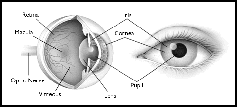
A cataract is a clouding of the normally clear lens of the eye. When the amount of light that passes through the lens is reduced and scattered by the cataract, images are not correctly focused on the retina at the back of the eye. The result is that vision becomes poor - it can be compared to looking through a frosted ors teamed window.
There are many misconceptions about cataract.
It is:
A cataract will often worsen to a point where surgery is needed to remove the cloudy lens and replace it with a permanent artificial lens.
Causes and Symptoms of Cataract
Causes and Cataract Development
Cataracts develop as a normal part of the ageing process. By the
age of 60 about half of all people will have some cataract formation although
it may be minor and not noticeable. By 70 years of age almost everyone
will have some degree of cataract formation. Other causes of cataract may
include family history, medical problems such as diabetes, injury to the
eye, medications such as steroids, various chronic eye diseases, and long-term,
unprotected exposure to sunlight.
Cataracts usually develop slowly and at a different rate in each
eye. The rate of cataract formation will also vary among individuals -
it is not possible to predict how fast cataracts will develop in any given
person. Most cataracts associated with ageing progress gradually over a
period of years.
Symptoms
Common symptoms include:
Detection
A thorough eye examination by an ophthalmologist
(eye doctor) can detect the presence and extent of a cataract, as well
as any other conditions that may be causing blurred vision or discomfort.
The examination will:
Surgery is the only way to remove the cataract.
This surgery may be necessary when vision has worsened to a point where
daily activities, reading and driving are affected, or if personal safety
is at risk. However if symptoms from a cataract are mild, a change of spectacles
may be all that is required. A decision to have a cataract removed should
be made only after a discussion with an ophthalmologist.
Cataracts cannot cannot be cured by any type of
medication, eye exercise, alternative therapy, diet or glasses.
Cataract Surgery and Intraocular Lenses
Success of Cataract Surgery
Removal of a cataract is the most common eye
operation and one of the most common surgical procedures performed in India.
It has a high rate of success due to the modern methods used by ophthalmologists.
If the eye is healthy, the likelihood is that cataract surgery will
restore good vision. Of every 100 operations to remove a cataract, 95 will
result in significantly improved vision. Despite the proven benefits of
cataract surgery, one should be aware that, like any surgery, there are
potential risks. Anyone considering cataract surgery should speak to their
ophthalmologist about this.
The lens is contained in a clear membrane called a capsule. With
older surgical methods both the lens and capsule were removed. With modern
methods the capsule is preserved. This is a significant advance in technique
because:
: the capsule can be used to hold and position the artificial lens
: the risks of surgery are fewer
: vision following surgery is usually better
Anaesthesia
cataract removal is usually performed under local
anaesthesia and light sedation. For some patients general anaesthesia may
be recommended. For those who have a local anaesthetic and sedation, they
may be able to see movement and light during the surgery but will not be
able to see the surgery.
Surgery
Ophthalmologists primarily use three different
methods to remove a cataract. All are performed with the ophthalmologist
viewing the eye through a microscope placed above the patient.
Phacoemulsification
This is the most common technique and usually takes from 20 to 60
minutes. A small incision of approximately 3mm is made where the cornea
meets the sclera (where the white of the eye meets the clear part). A small
probe which vibrates at high frequencies is inserted into the eye to divide
the cloudy lens into small pieces. The pieces are then suctioned away leaving
only the lens capsule.
An artificial lens is then usually inserted into the lens capsule.
The incision is usually so small that often stitches are not required.
Extracapsular Extraction
This technique is used less commonly but it is effective for those
patients whose lens is too hard to remove using phacoemulsification. A
10 to 12mm incision is made whre the sclera meets the cornea. The front
of the capsule is opened and the lens then removed. An artificial lens
is inserted and fixed into position. The incision is then closed with several
sutures.
Intracapsular Extraction
The entire lens and capsule is removed. This is now only used in
special cases.And still many part of India.
Artificial Lenses
This is also called an "intraocular lens" or an "IOL". It is a transparent
plastic disk with a similar shape to a natural human lens. The focus of
the lens is fixed and cannot change. The strength of the lens chosen for
an individual is determined by the ophthalmologist's prescription and made
for distance vision.
Most people fitted with IOLs will not need spectacles for distance
vision. However, other may require glasses for both distance vision and
close vision, such as reading. The use of intraocular lenses has almost
eliminated the need to thick cataract glasses and contact lenses.
Recovery from Surgery
if the operation has been performed under local
anaesthetic, after surgery the patient is moved to a quiet area to recover
from the effects of sedation. Most patients are then ready to leave fro
home within a few hours. Most patients get sufficient pain relief by using
over-the-counter products such as paracetamol. Some patients may require
more pain relief and are encouraged to see their ophthalmologist.
Within a number of days the ophthalmologist will examine the eye.
Recovery is usually quick. Soon after surgery most people notice their
vision has improved although glasses are still needed for reading. If the
operation has been an extracapsular extraction then inflammation from the
operation may take several days or weeks to setle down. Vision may remain
hazy or cloudy during that time but slowly improve over the next three
months.
Typically lights will appear intense shortly after the operation
and during this time sunglasses may be worn.
After the eye has healed and adjusted to the intraocular lens for
several weeks the eye is tested again and a new prescription for glasses
may be needed.
